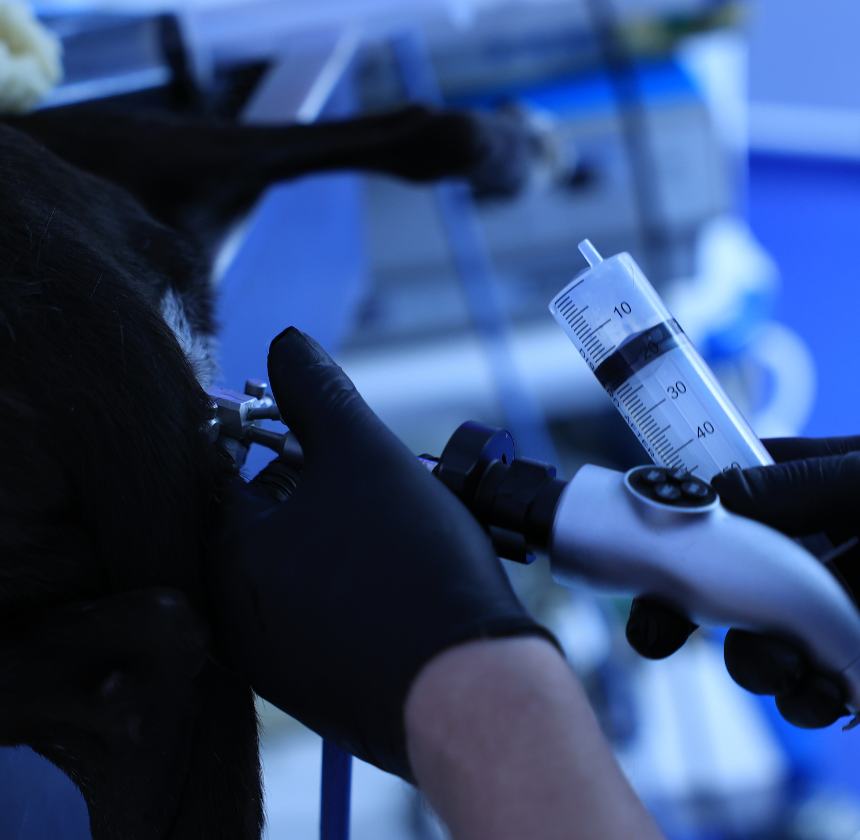
Vaginoscopy
We offer state-of-the-art veterinary vaginoscopy, a specialised procedure designed to examine the vaginal canal and reproductive tract of female animals with precision and accuracy.
Conditions Diagnosed & Treated with Vaginoscopy
Vaginoscopy is a diagnostic procedure performed by veterinarians to examine the vaginal canal of female animals. It involves the use of a specialised tool called a vaginoscope, which is a slender, flexible tube equipped with a light source and a camera. With the vaginoscope, we can obtain clear and detailed images of the internal structures of the vagina, cervix, and sometimes the uterus.
Vaginitis
Inflammation of the vaginal lining, which can be caused by infections, irritants, or allergies.
Vaginal Discharge
Vaginoscopy can help identify the cause of abnormal vaginal discharge, such as infection, inflammation, or the presence of foreign bodies.
Vaginal Tumours & Masses
The procedure allows for visualisation of any abnormal growths or lesions within the vaginal canal.
Cervical Abnormalities
Vaginoscopy can be used to evaluate the cervix for abnormalities such as strictures, polyps, or neoplasia.
Foreign Bodies
Vaginoscopy can help identify and locate foreign objects lodged within the vaginal canal, such as grass seeds or other debris.
Reproductive Tract Infections
Various infections can affect the reproductive tract, including the vagina, cervix, and uterus. Vaginoscopy assists in identifying the site and nature of infection, aiding in targeted treatment.
Reproductive Disorders
Conditions such as cysts, polyps, tumours, or strictures within the reproductive tract can be visualised through vaginoscopy. This aids in diagnosing the nature and extent of the disorder and guiding appropriate management or surgical intervention.
Bleeding Evaluation
Vaginoscopy is sometimes used as part of a breeding soundness evaluation in female animals. It helps assess the readiness of the reproductive tract for breeding, detect any abnormalities that may affect fertility, and evaluate post-breeding responses.
Reproductive Trauma
Trauma to the reproductive tract, including the vagina, may occur due to injuries, difficult births, or medical interventions. Vaginoscopy can assess the extent of trauma and guide treatment decisions.
Vaginoscopy Procedure
Vaginoscopy procedure involves several steps to safely and effectively visualise the vaginal canal and surrounding reproductive structures in female animals. Here is an overview of the typical process.
Pre-procedure Preparation
Before the procedure, we will conduct a thorough physical examination of the animal to assess overall health and suitability for anaesthesia. Pre-operative blood work may also be performed to evaluate organ function and ensure the pet’s ability to tolerate anaesthesia.
Anaesthesia or Sedation
Vaginoscopy is usually performed under general anaesthesia to ensure the pet remains still and comfortable throughout the procedure. We will administer an appropriate anaesthetic agent based on the pet’s size, age, and health status.
Vaginoscopy Procedure
Once the pet is under anaesthesia, they will be positioned appropriately for the vaginoscopy procedure. This may involve placing the animal in dorsal recumbency (on their back) or lateral recumbency (on their side), depending on the size and anatomy of your pet.
A specialised vaginoscope, which is a slender, flexible tube equipped with a light source and camera, is carefully inserted into the vaginal canal. The vaginoscope may be lubricated to ease insertion and minimise discomfort for the animal.
As the vaginoscope is advanced through the vaginal canal, we will observe the images transmitted by the camera on a monitor. This allows for real-time visualisation of the vaginal walls, cervix, and surrounding structures.
We will thoroughly examine the vaginal canal and reproductive structures for any abnormalities, such as inflammation, masses, or foreign bodies. If suspicious lesions are identified, we may collect biopsy samples for further evaluation.
Post-Procedure Care
Once the vaginoscopy procedure is complete, your pet will be carefully monitored as they recover from anaesthesia. They may be kept in a warm, quiet recovery area until they are fully awake and alert. Postoperative pain management and supportive care may be provided as needed.
Vaginoscopy Benefits in Pets
Overall, veterinary vaginoscopy is a valuable diagnostic tool that allows us to evaluate and manage various reproductive and vaginal conditions in female animals with precision and accuracy.
Accurate Diagnosis
Allows direct visualisation of the vaginal canal to identify abnormalities like infections, strictures, tumours, or injuries.
Minimally Invasive
Reduces the need for exploratory surgery, minimising discomfort and recovery time.
Targeted Sampling
Enables precise collection of samples for diagnosing infections, hormonal conditions, or other issues.
Foreign Body Removal
Safely retrieves objects without the need for surgery.
Breeding Assessments
Assists in evaluating reproductive health and identifying anatomical issues that may affect breeding.
Early Detection
Helps detect issues like vaginal infections or structural abnormalities before they worsen.

