Thoracoscopy
We understand the concern and care you have for your furry companions, especially when it comes to their respiratory health.
Conditions Diagnosed & Treated with Thoracoscopy
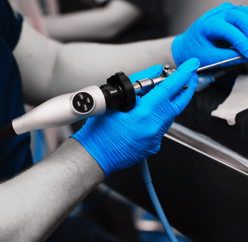
Thoracoscopy is an advanced diagnostic and therapeutic technique that we offer to help diagnose and treat a range of respiratory conditions in pets with precision and minimal invasiveness. With the use of a specialised instrument called a thoracoscope, which is a thin, flexible tube equipped with a camera and light source, we can visualise and access the thoracic cavity, the space within the chest that houses the lungs and other vital organs.
Pleural Effusion
Thoracoscopy can help diagnose the cause of pleural effusion (accumulation of fluid in the chest cavity), such as infections, heart disease, cancer, or trauma. It also allows for the drainage of excessive fluid and sampling for further analysis.
Pneumothorax
This condition occurs when air accumulates in the pleural space, causing lung collapse. Thoracoscopy can be used to locate the source of air leakage and perform corrective procedures such as sealing air leaks or removing trapped air.
Lung Tumours
Thoracoscopy enables visualization of lung tissue and identification of tumors or abnormal growths. Biopsy samples can be obtained for histopathological examination to determine the nature of the tumor and guide treatment decisions.
Foreign Body Removal
localisationPets may inhale or ingest foreign objects that become lodged in the airways or chest cavity. Thoracoscopy allows for precise localization and minimally invasive removal of foreign bodies, reducing the need for extensive surgery.
Mediastinal Masses
Masses or tumoyrs in the mediastinum (the space between the lungs) can cause respiratory distress and other symptoms. Thoracoscopy can aid in the biopsy and removal of mediastinal masses, improving diagnostic accuracy and treatment options.
Diaphragmatic Hernia
In cases of traumatic diaphragmatic hernia, where abdominal organs protrude into the chest cavity, thoracoscopy can assist in visualizing the hernia defect and repairing it surgically with minimal invasiveness.
Chronic Respiratory Conditions
Thoracoscopy may be indicated in pets with chronic respiratory conditions such as chronic bronchitis, interstitial lung disease, or recurrent pneumonia to assess lung tissue, collect samples for analysis, and guide treatment strategies.
Lube Lobe Torsion
Thoracoscopy can be used to investigate and potentially correct lung lobe torsion, a condition where a portion of the lung twists upon itself, leading to compromised blood flow and respiratory function.
Thoracoscopy Procedure
The procedure for performing thoracoscopy on pets involves several steps and requires specialised equipment. Here’s an overview of the typical procedure:
Pre-procedure Preparation
Before the procedure, we will conduct a thorough physical examination of your pet and review relevant medical history and diagnostic imaging results. The pet may need to undergo preoperative blood tests and imaging studies (such as X-rays or ultrasound) to evaluate the extent of the condition and plan the procedure accordingly.
Anaesthesia or Sedation
Thoracoscopy in pets requires general anaesthesia to ensure the animal remains still and comfortable throughout the procedure. We will select an appropriate anaesthetic protocol based on the pet’s health status, age, and other factors. Intravenous catheters may be placed to administer medications and fluids during the procedure.
Thoracoscopy Procedure
Once the pet is anaesthetised, they will be positioned on their back or side on the surgical table, exposing the chest area for the procedure. The area around the incision sites will be shaved and prepared for aseptic surgery.
Small incisions, typically ranging from 5mm to 15mm in size, are made in the chest wall using a scalpel or trocar under sterile conditions. These incisions serve as entry ports for the thoracoscope (a thin, flexible tube with a camera and light source) and surgical instruments.
The thoracoscope is carefully inserted through one of the incisions into the thoracic cavity. The camera at the tip of the thoracoscope provides real-time high-definition images of the internal structures, allowing the veterinarian to navigate and visualise the lungs, pleura, mediastinum, and other thoracic organs.
We will then systematically examine the thoracic cavity, looking for abnormalities such as tumours, infections, fluid accumulation (pleural effusion), foreign bodies, or diaphragmatic hernias. Biopsy samples, aspirates, or fluid may be collected for laboratory analysis to aid in diagnosis.
Post-Procedure Care
Your pet is carefully monitored as they recover from anaesthesia. They may be placed in a warm, quiet recovery area until fully awake. Postoperative pain management and supportive care are provided as needed to ensure the pet’s comfort and well-being.
Thoracoscopy Benefits in Pets
Overall, veterinary thoracoscopy provides a safer, less invasive, and more effective approach to diagnosing and treating thoracic conditions in pets, ultimately improving their quality of life and well-being.
Accurate Diagnosis
Provides a clear view of the chest cavity to identify conditions like tumours, infections, or fluid buildup.
Minimally Invasive
Offers a safer alternative to open chest surgery, resulting in less pain and quicker recovery for pets.
Precise Biopsies
Allows targeted collection of tissue samples for diagnosing lung, pleural, or mediastinal diseases.
Therapeutic Applications
Facilitates procedures such as removing masses, draining fluid, or treating pneumothorax directly.
Early Intervention
Detects thoracic diseases at earlier stages for prompt and effective treatment.
Reduced Recovery Time
Minimises surgical trauma, leading to faster healing and less postoperative discomfort.

