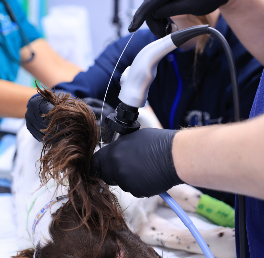
Video Otoscopy
As part of our commitment to excellence we are pleased to offer state-of-the-art otoscopy services for our furry companions.
Conditions Diagnosed & Treated with Otoscopy
Pet Video Otoscopy refers to the use of an otoscope or endoscope to examine the ear canal and tympanic membrane of a pet, such as a dog or cat. This procedure is commonly performed by veterinarians to diagnose and treat various ear conditions, including infections, foreign bodies, tumours, and ear mites.
Otitis Externa
This is inflammation or infection of the external ear canal. Otoscopy helps identify the cause of the inflammation, such as bacteria, yeast, or foreign bodies, and allows for targeted treatment.
Otitis Media
Inflammation or infection of the middle ear. Otoscopy can help visualise the tympanic membrane and assess the extent of the infection.
Ear mites (Otodectes cynotis)
These are parasitic mites that infest the ear canal, causing irritation and inflammation. Otoscopy allows for direct visualisation of the mites and their effects on the ear canal.
Foreign Bodies
Objects such as grass seeds, foxtails, or other debris can become lodged in the ear canal, leading to irritation, infection, and discomfort. Otoscopy helps locate and remove these foreign bodies safely.
Tumours
Otoscopy can aid in the detection of tumors or abnormal growths within the ear canal or on the tympanic membrane. Early detection allows for prompt treatment and management.
Ear Polyps
These are benign growths that can develop in the ear canal or middle ear. Otoscopy helps visualise the size and location of the polyps for appropriate treatment planning.
Chronic Otitis
Some pets may suffer from recurrent or persistent ear infections. Otoscopy can help identify underlying causes, such as anatomical abnormalities or allergic reactions, to develop a comprehensive treatment approach.
Ear Trauma
Injuries to the ear canal or tympanic membrane can occur due to trauma or accidents. Otoscopy helps assess the extent of the damage and guide appropriate treatment.
Video Otoscopy Procedure
The procedure for performing otoscopy on pets involves several steps and requires specialised equipment. Here’s an overview of the typical procedure.
Pre-procedure Preparation
Our veterinarian usually examines your pet to assess its overall health and determine if an otoscopy is necessary. To reduce the risk of complications during anaesthesia, your pet may be fasted for a certain period beforehand.
Anaesthesia or Sedation
Otoscopy in pets typically requires general anaesthesia to ensure the pet remains still and comfortable throughout the procedure. We will carefully select an appropriate anaesthetic protocol based on your pet’s age, breed, health status, and the nature of the otoscopy.
Video Otoscopy Procedure
We will gently insert a small, flexible tube with a light and camera attached (endoscope) into the ear canal. The endoscope allows for direct visualisation of the ear structures on a monitor.
We carefully navigate the endoscope through the ear canal, examining the ear structures, including the ear canal walls, tympanic membrane (eardrum), and any abnormalities present. We may manipulate the endoscope to get a comprehensive view of the entire ear canal.
During the examination, we assess the condition of the ear, noting any signs of inflammation, infection, foreign bodies, tumours, or other abnormalities. The findings are documented for further evaluation and treatment planning.
If necessary, we may collect samples from the ear canal for further analysis. This may include swabs for cytology (microscopic examination of cells) or culture and sensitivity testing to identify the underlying cause of infection and guide antibiotic therapy.
Depending on the findings of the examination, we may administer treatment immediately following otoendoscopy. This may include ear cleaning, medication administration (e.g., antibiotics, antifungals), removal of foreign bodies or debris, or other interventions tailored to the specific condition of the pet.
Post-Procedure Care
After the procedure, the pet is closely monitored as it recovers from sedation or anaesthesia. We will provide post-procedure instructions for at-home care, such as administering medications or monitoring for any signs of complications.
Video Otoscopy Benefits in Pets
Overall, video otoscopy offers significant advantages for pets by providing us with a comprehensive view of the ear structures, facilitating accurate diagnosis, and enabling tailored treatment plans. This leads to improved outcomes and better quality of life for pets with ear-related problems.
Accurate Diagnosis
Provides a detailed view of the ear canal and eardrum, helping identify infections, foreign bodies, polyps, or tumours.
Minimally Invasive
Offers a safer and less invasive alternative to traditional methods for ear examination and treatment.
Enhanced Cleaning
Enables thorough removal of wax, debris, or infected material from the ear canal.
Improved Treatment
Allows precise delivery of medication or irrigation to affected areas.
Early Detection
Detects ear issues, such as infections or blockages, before they worsen.
Surgical Assistance
Aids in guiding procedures like removing polyps or foreign objects without the need for extensive surgery.

