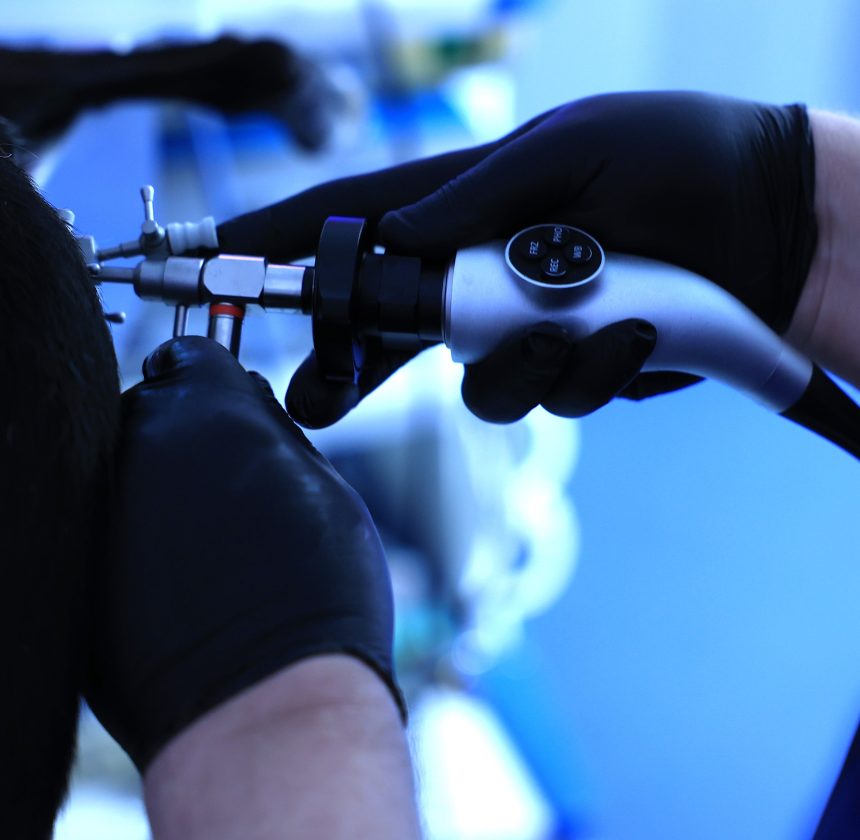
Cystoscopy
We offer veterinary urethrocystoscopy, simply known as cystoscopy, a specialised procedure designed to examine the urinary tract, bladder, and urethra.
Conditions Diagnosed & Treated with Cystoscopy
Cystoscopy is a minimally invasive procedure used to examine the urinary tract of animals, including the bladder and urethra. Similar to the human procedure, a cystoscope—a slender, flexible, or rigid tube equipped with a camera and light—is inserted into the urinary opening and gently guided through the urethra into the bladder.
Bladder Stones
Cystoscopy allows us to directly visualise bladder stones (uroliths) and assess their size, number, and location within the bladder. This information helps guide treatment decisions, such as whether surgical removal or non-surgical dissolution is necessary.
Bladder Tumours
Cystoscopy aids in the detection and evaluation of bladder tumours in animals. By visualising the bladder lining, we can identify suspicious lesions, take biopsies for further analysis, and determine the extent of tumour involvement.
Bladder Inflammation (Cystitis)
Inflammatory conditions of the bladder, such as cystitis, can cause discomfort and urinary symptoms in animals. Cystoscopy allows us to assess the degree of inflammation, identify potential causes (such as infection or bladder stones), and develop appropriate treatment strategies.
Urethral Obstructions
Cystoscopy can help diagnose and evaluate urethral obstructions, which may occur due to urinary stones, tumours, or anatomical abnormalities. By visualising the urethra, we can identify the site and nature of the obstruction and determine the best course of action for relieving it.
Congenital Anomalies
Some pets may be born with congenital abnormalities of the urinary tract, such as urethral strictures or ectopic ureters. Cystoscopy enables us to visualise these anomalies directly and plan appropriate treatment strategies, which may include surgical correction.
Urinary Tract Infections (UTIs)
While cystoscopy itself is not typically used to diagnose urinary tract infections, it can be helpful in cases where UTIs are recurrent or complicated. By visualising the bladder lining and collecting urine samples for culture, we can identify potential sources of infection and guide treatment decisions.
Haematuria (Blood in the Urine)
Pets with haematuria (blood in the urine) may undergo cystoscopy to determine the underlying cause. Visual inspection of the bladder and urinary tract can help identify sources of bleeding, such as bladder tumours, inflammation, or trauma
Cystoscopy Procedure
Cystoscopy involves several steps to visualise and evaluate the urinary tract of pets. Here is an overview of the typical veterinary cystoscopy procedure.
Pre-procedure Preparation
Before the procedure, your pet may undergo preoperative assessments, including blood tests and imaging studies, to evaluate its overall health and identify any underlying conditions. If necessary, the animal may also receive preoperative medications for sedation or anaesthesia.
Anaesthesia or Sedation
Cystoscopy is usually performed under general anaesthesia to ensure the pet remains still and comfortable throughout the procedure. We will administer an appropriate anaesthetic agent based on the pet’s size, age, and health status.
Cystoscopy Procedure
Once the animal is anaesthetised, it is positioned appropriately for the cystoscopy procedure. This may involve placing the animal on its back or side to allow easy access to the perineal region.
The cystoscope, a specialised instrument with a light source and camera, is prepared for use. Depending on the procedure’s specific requirements and the animal’s size, the cystoscope may be a flexible or rigid tube.
We gently insert the cystoscope into the animal’s urinary opening (urethra) and advance it slowly into the bladder. The cystoscope is carefully navigated through the urinary tract, allowing for visualisation of the bladder lining and urethra.
As the cystoscope is advanced, real-time images of the urinary tract are transmitted to a monitor, allowing us to observe the internal structures of the bladder and urethra. We may manipulate the cystoscope to obtain different views and thoroughly inspect the entire urinary tract.
During cystoscopy, we evaluate the bladder and urethra for abnormalities such as stones, tumours, inflammation, or anatomical defects. If suspicious lesions are identified, biopsies may be taken for further analysis to confirm diagnoses
Post-Procedure Care
After the cystoscopy procedure is completed, the animal is carefully monitored as it recovers from anesthesia. Postoperative pain management and supportive care may be provided as needed to ensure the animal’s comfort and well-being.
Cystoscopy Benefits in Pets
Overall, veterinary cystoscopy is a valuable diagnostic and therapeutic procedure that allows us to visualise and evaluate the urinary tract of animals, leading to accurate diagnoses and appropriate treatment plans.
Accurate Diagnosis
Provides a direct view of the urinary bladder and urethra to identify stones, tumours, inflammation, or structural abnormalities.
Minimally Invasive
Avoids the need for open surgery, reducing recovery time and discomfort.
Targeted Sampling
Enables precise collection of urine, tissue, or stone samples for diagnosis.
Foreign Body Removal
Safely retrieves objects or bladder stones without requiring invasive procedures.
Therapeutic Applications
Assist in treating conditions like strictures or urethral obstructions.
Early Detection
Identifies urinary tract diseases at an earlier stage for timely and effective treatment.

