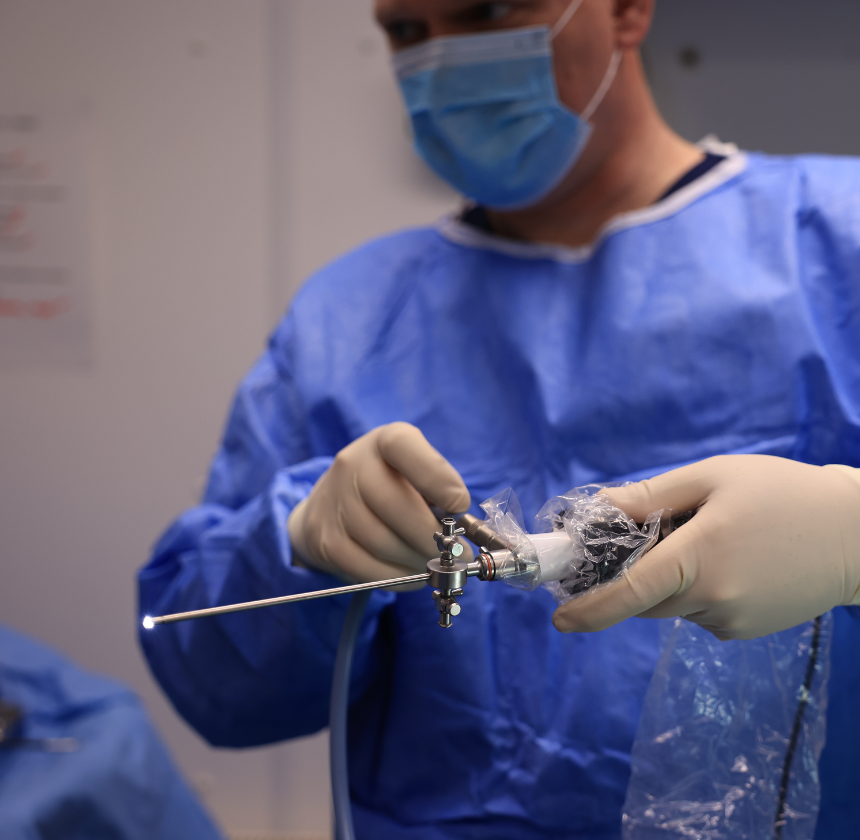
Arthroscopy
We offer veterinary arthroscopy, a minimally invasive procedure to diagnose and treat joint conditions, providing detailed insights and targeted treatments with small incisions.
Conditions Diagnosed & Treated with Arthroscopy
Arthroscopy is a minimally invasive procedure used to diagnose and treat joint conditions in pets. Using small incisions and a specialised camera (arthroscope), we can view the inside of your pet’s joints with precision, allowing us to assess their condition and perform targeted treatments without the need for large incisions or extensive surgery.
Arthroscopy is commonly used for joint issues in the shoulder, elbow, knee, and hip. This technique allows us to identify problems such as cartilage damage, joint instability, and inflammation, which may not be visible on X-rays or traditional imaging
Joint Pain or Limping
When your pet experiences persistent joint pain or limping, and standard diagnostic methods (like X-rays) fail to identify the issue, arthroscopy provides a detailed, real-time view of the joint structures to pinpoint the source of discomfort.
Cruciate Ligament Injuries
Arthroscopy is commonly used to assess and treat damage to the cruciate ligaments, which are vital for knee stability. It allows for accurate identification of tears or sprains and offers the ability to repair them with minimal tissue disruption.
Osteoarthritis
For pets with chronic arthritis or joint inflammation, arthroscopy allows for precise evaluation of the joint surfaces, including cartilage wear and synovial membrane condition, aiding in effective treatment options, such as debridement or smoothing of damaged cartilage.
Damaged Cartilage
Arthroscopy can identify and treat damage to joint cartilage or bone that might not be visible on conventional X-rays, enabling surgical interventions to repair or remove damaged tissue, promoting better joint function.
Joint Infections or Swelling
When a joint becomes infected or swollen without a clear cause, arthroscopy allows for direct visualisation of the joint cavity, enabling the collection of fluid or tissue samples for diagnosis and facilitating the removal of infected material if necessary.
Abnormalities in Joint Structures
In cases where the joint structures are suspected to be abnormal due to congenital conditions or trauma, arthroscopy provides an in-depth view to identify structural issues such as malalignment, loose bodies, or other deformities, offering the opportunity for corrective treatment.
Arthroscopy Procedure
Arthroscopy involves several key steps to visualise and treat joint conditions in pets. Here is an overview of the typical veterinary arthroscopy procedure.
Pre-procedure Preparation
Before the procedure, your pet will undergo a physical examination, blood tests, and possibly imaging such as X-rays or CT scans to assess the condition of the joint. This helps us determine if arthroscopy is the best approach.
Anaesthesia or Sedation
Arthroscopy is performed under general anesthesia to ensure your pet remains comfortable and still throughout the procedure. An intravenous catheter will be placed for fluid administration and medications.
Arthroscopy Procedure
The procedure begins with small incisions (usually 2-3) over the joint. A sterile saline solution is used to fill the joint space, allowing the camera (arthroscope) to be inserted and providing a clear view. The arthroscope is connected to a monitor that displays high-definition images of the joint.
Assessment and Treatment
Once the joint is visualised, we will inspect the structures, such as the cartilage, ligaments, and synovial lining, for any damage or abnormalities. If necessary, surgical instruments can be inserted through additional small incisions to remove debris, repair damaged cartilage, or perform other treatments. This may include:
- Debridement: Cleaning up torn or damaged tissue.
- Cartilage repair: smoothing or repairing damaged cartilage surfaces.
- Foreign body removal: removing foreign materials that may be causing pain or inflammation.
Closure and Recovery
Once the procedure is complete, the incisions are closed with sutures or surgical glue. Since arthroscopy involves minimal tissue disruption, pets typically experience less pain and discomfort. Recovery times are generally quicker compared to traditional surgery.
Post-Procedure Care
After arthroscopy, your pet will be monitored in a recovery area as they wake from anesthesia. Pain management is an important part of recovery, and medications will be prescribed to ensure comfort. Activity restrictions will be advised to allow proper healing of the joint, and follow-up appointments may be scheduled to assess recovery progress.
Most pets recover within a few days to weeks, depending on the complexity of the condition treated and the specific joint involved. The majority of pets return to normal activity after a period of rehabilitation.
Arthroscopy Benefits in Pets
Overall, veterinary arthroscopy is a valuable diagnostic and therapeutic procedure that allows us to visualise and evaluate the internal structures of joints, leading to accurate diagnoses and effective treatment plans for your pet.
Accurate Diagnosis
The camera allows us to visualise the inside of the joint, providing a clear and detailed picture of the issue.
Minimally Invasive
Smaller incisions reduce trauma to surrounding tissues, resulting in less pain and a faster recovery.
Faster Recovery
Pets typically experience less pain and quicker recovery times compared to conventional surgery.
Targeted Treatment
With arthroscopy, we can perform precise treatments, such as removing damaged tissue or repairing cartilage, without the need for large surgical openings.
Reduced Risk of Infection
Smaller incisions lower the risk of infection and other complications.

